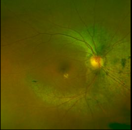
Case 10: Optomap® plus with Resmax™ Color Image OD

Case 10: A 64 yo BF presented for follow up of peri-central RP. Patient has a history of diabetes and hypertension. She has undergone focal laser for the treatment of diabetic retinopathy in her left eye. Patient reported no change in vision since her laser surgery 1 year ago. BCVA was 20/25+2 OD and 20/40+2 OS. Examination of the posterior pole revealed pigmentary changes following the arcades in both eyes with macular sparing, consistent with the stable VAs. Lipid deposition was found in the macular area and is most likely secondary to diabetes or hypertension. Flash ERGs revealed a decreased response under all conditions with a greater reduction under cone, rather than rod, conditions.


