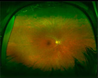
Case 1 Optomap® Color Fundus Image OD

Case 1: A 36 yo WF presented for follow-up of long standing RP. Patient reported that she has had trouble seeing at night since her mid teens. Though she only reported visual field loss 18 months earlier, the patient noted that she had a history of bumping into things which she attributed to clumsiness. BCVA was 20/20 OD and OS. Retina evaluation revealed mid-peripheral bone spicules and arterial attenuation in both eyes. Corresponding to the posterior pole examination, previous FDT-30 degree fields revealed a ring scotoma and more current Goldmann fields revealed constriction in all quadrants. The patient had previously undergone testing for autosomal dominant RP which failed to reveal any mutations. The patient has agreed to pursue testing for other RP genetic variations.


