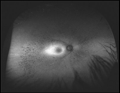
Case 1: Optomap® FAF Image OD

AF of the right eye reveals scattered areas of hypo-AF in the mid and far periphery which are due to dead RPE cell and RPE migration and hence no lipofuscin (LF) accumulation. The macula reveals a ring of hyper-AF which is due to “sick” RPE cells and increased LF and suggests that the condition is still progressing. In time hyper-AF areas become zones of hypo-AF.


