

Topcon 3D OCT OD
Green arrow depicts a well defined Bruch’s Membrane (BM). Note the
extreme thinning of the overlying RPE and absence of the PIL. BM is
masked in normal eyes because of the presence of a normal RPE.

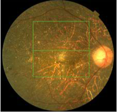
The choroidal vasculature is
observable above because
much of the overlying RPE and
choriocapillaris have been lost.
Small, discrete white dots
are localized to the anterior
border of BM with the aid of SD OCT.
The arterioles are quite attenuated.