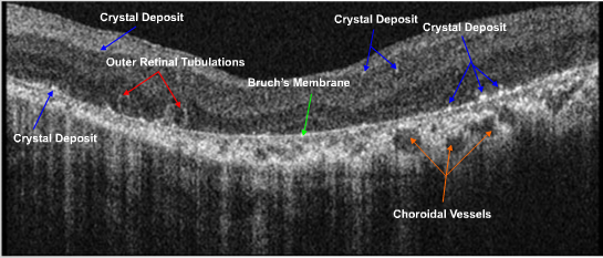
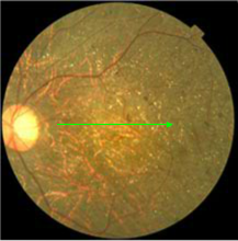
Topcon 3D OCT OS
The fundus appearance is similar OS. In addition to the white dots on BM, multiple hollow circular bodies are found In the SD OCT cross sectional images. Large choroidal vessels are also visualized and are unmasked because of the loss of overlying tissue.


