Case #1 – Pathological Myopia

Pathological Myopia: Mini to Maxi Bulge – Page 9 of 19

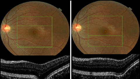
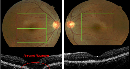
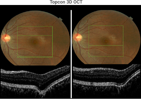
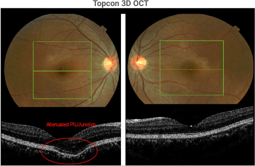
Diagnosis – sub-clinical mini-bulges secondary to myopiaTreatment - Since some authorities believe that IOPs can result in posterior staphyloma in patients with genetic weak elements of the sclera, diurnal IOPs are advisable. Treatment of patients with high IOPs or spikes in IOPs should be considered to prevent loss of the retinal nerve fiber layer (RNFL)

Pathological Myopia: Mini to Maxi Bulge – Page 9 of 192020-08-24T17:36:43+00:00

Pathological Myopia: Mini to Maxi Bulge – Page 5 of 19

Topcon 3D OCT 2 dimensional video which is composed of 128 horizontal sections beginning from the top and going down. All sections are contained within the green box on the previous page. Note: The movies on page 5 & 6 can easily be paused or replayed by moving the mouse

Pathological Myopia: Mini to Maxi Bulge – Page 5 of 192020-06-30T13:41:18+00:00

Pathological Myopia: Mini to Maxi Bulge – Page 4 of 19

A review of the 128 horizontal sections through the left posterior pole reveals two mini-bulges, one superior to the fovea (above left) and one inferior to the fovea (above right). The most likely interpretation of these two mini-bulges is that they represent very early scleral weakening that could lead to a clinically evident posterior

Pathological Myopia: Mini to Maxi Bulge – Page 4 of 192020-06-02T10:47:18+00:00

Pathological Myopia: Mini to Maxi Bulge – Page 3 of 19

The section through the fovea in the right eye demonstrates the mini-bulge just nasal to the fovea in the image on the left. The PIL/junction (see page 10) is still present but attenuated. The section through the fovea in the left eye is normal. The bulge is likely due to a localized weakness in the

Pathological Myopia: Mini to Maxi Bulge – Page 3 of 192020-06-02T10:46:03+00:00

Pathological Myopia: Mini to Maxi Bulge – Page 2 of 19

History / Chief complaintAsymptomatic 22 year old Asian female with gradual increase in myopia in both eyes over the last 5 years.Clinical FindingsBCVA: OD 20/20 and OS 20/20 with  -7.50 OUDFE: Unremarkable with no evidence of myopic changes

Pathological Myopia: Mini to Maxi Bulge – Page 2 of 192020-08-24T16:26:51+00:00

Pathological Myopia: Mini to Maxi Bulge – Page 1 of 19

Pathological Myopia deserves our attention and recognition as the seventh ranking cause of legal blindness in the US and even ranks higher in many Asian nations. The increased axial length is due in part to genetically weak areas of the sclera resulting in thinning and stretching. Optos Ultra-Widefield 3D Wrap depicts many of the

Pathological Myopia: Mini to Maxi Bulge – Page 1 of 192020-06-24T05:55:43+00:00