

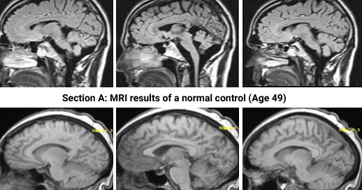
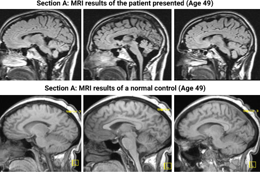
Non-glaucomatous RNFL loss with Cerebellar Atrophy – Page 31 of 31
References M.E Hajee, W.F March, D.R Lazzaro, A.H Wollintz, E.M Shrier, S. Glazman, and I.G Bodis-Wollner. 2009 Inner Retinal LayerThinning in Parkinson's Disease. Archives of Ophthalmology; 127 (6) 737-741 K. Agan, D. Kutlu, N. Basak, O. Us, and D. ince-Gönal. 2006 Spinocerebellar Ataxia Type 2 In A Turkish Family. Marmara Medical Journal; 19 (3)135-138