

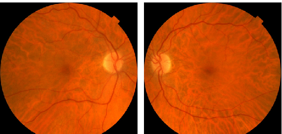
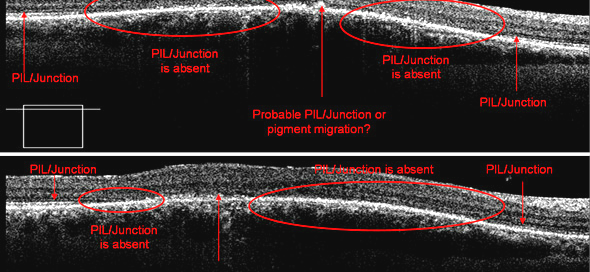
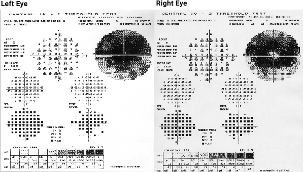
Plaquenil Induced Retinal Toxicity – Page 16 of 25
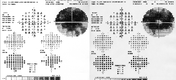
Top images demonstrate the actual mfERGs in one specific normal subject. The bottom images compare these results to the normative database. The central hill in the images left (top images) from a normal subject demonstrates increased sensitivity of the macula. According to the color code in the images below, the normal subject varies very