Case #5 – Vein Occlusion

Three Quadrant Retinal Vein Occlusion – Page 19 of 19

References: Ota M„ Tsujikawa A, Kita M, Miyamoto K, Sakamoto A, Yamaike N, Kotera Y, Yoshimura N. Integrity of Foveal Photoreceptor Layer in Central Retinal Vein Occlusion. The Journal of Retinal and Vitreous Diseases; 2008:28(10): 1502-1508 H Koizumi, D. C. Ferrara, C. Brue, and R. F. Spaide. Central Retinal Vein Occlusion Case- Control Study. American

Three Quadrant Retinal Vein Occlusion – Page 19 of 192021-11-22T23:08:52+00:00

Three Quadrant Retinal Vein Occlusion – Page 17 of 19

Jerome Sherman, OD, FAAO Jerome Sherman, O.D., is perhaps optometry's most prolific writer, publishing over 650 clinical articles, research manuscripts, book chapters and two CDs. He is senior author of three books that were published in 2007, and has delivered over three thousand lectures both nationally and internationally. He has served as a contributing

Three Quadrant Retinal Vein Occlusion – Page 17 of 192021-11-22T23:08:25+00:00

Three Quadrant Retinal Vein Occlusion – Page 16 of 19

Possible Contributory Etiologies of RVO Activated Protein C Resistance 5 Benign Retinal Vasculitis 3 Arteriovenous crossing abnormality (BRVO) 5 Presumed Phlebitis of the optic disc vessels 5 Hypertension 2,3 Diabetes Mellitus 2,3 Kidney Disease 3 Older Age 3 Urban Location 3 Hyperlipidaemia 5 Atherosclerosis 5 Glaucoma 2,3 Atrial Fibrillation

Three Quadrant Retinal Vein Occlusion – Page 16 of 192021-11-22T23:08:16+00:00

Three Quadrant Retinal Vein Occlusion – Page 15 of 19

Suggested Laboratory Studies in Retinal Vein Occlusion Complete Blood Count Erythrocyte Sedimentation Rate Platelet Count Prothrombin Time Activated Partial Thromboplastin Time Bleeding time Lipid profile Plasma Homocysteine Plasma Fibrinogen D-dimer Fibrinogen Degradation Products Antithrombin III Protein S Protein C Antinuclear Antibodies Anticardiolipin Antibodies Lupus Anticoagulant Venereal Disease Research Laboratory (VDRL)

Three Quadrant Retinal Vein Occlusion – Page 15 of 192021-11-22T23:08:07+00:00

Three Quadrant Retinal Vein Occlusion – Page 14 of 19

The following is a list of links to the companies and contributors of the Three Quadrant Retinal Vein Occlusion: http://www.lulu.com/content/hardcover-book/the-retinal-nerve-fiber-layer-in-clinical-practice/1775291 http://www.lulu.com/content/hardcover-book/photoreceptor-integrity-line-as-revealed-by-spectral-domain-oct/5738467

Three Quadrant Retinal Vein Occlusion – Page 14 of 192021-11-22T23:07:57+00:00

Three Quadrant Retinal Vein Occlusion – Page 13 of 19

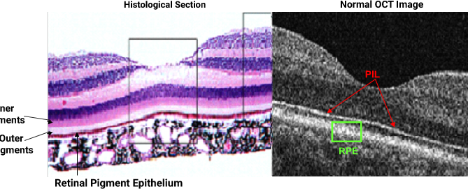
Histological Section as Compared to the OCT ImageAlthough the photoreceptor integrity line, or the PIL (defined as the junction between the inner and outer segments) is barely visible in most histological sections, it is highly prominent in normal SD OCTs. The PIL, as shown above, should be continuous throughout the entire scan in normal eyes.

Three Quadrant Retinal Vein Occlusion – Page 13 of 192021-11-22T23:07:48+00:00

Three Quadrant Retinal Vein Occlusion – Page 12 of 19

Comments and Conclusions Most patients with vein occlusions are elderly with systemic disorders such as hypertension and diabetes.2,3,4 In contrast, our patient is young, trim and fit, and without any identifiable systemic disorder. More than two decades earlier at the age of 30, he presented with a CRVO as a young, healthy adult, a

Three Quadrant Retinal Vein Occlusion – Page 12 of 192021-11-22T23:07:39+00:00

Three Quadrant Retinal Vein Occlusion – Page 11 of 19

We Introduce SLT to our Patients by Using the Following Animation powered by Eyemaginations.com Note: White arrow denotes the trabecular meshwork The newest surgical option for the treatment of open angle glaucoma is called selective laser trabeculoplasty, or SLT. A low frequency laser beam is directed into the trabecular meshwork, which is

Three Quadrant Retinal Vein Occlusion – Page 11 of 192021-11-22T23:07:31+00:00

Three Quadrant Retinal Vein Occlusion – Page 10 of 19

Retinal nerve fiber layer abnormalities OU as shown with GDx. The Deviation Map reveals an inferior loss OU which has progressed somewhat OD. Both the fields and the GDx shown were obtained prior to the recent vein occlusion. Learn more about GDx and OCT RNFL interpretation.* *The Retinal Nerve Fiber Layer in Clinical

Three Quadrant Retinal Vein Occlusion – Page 10 of 192021-11-22T23:07:23+00:00