Case #9 – AV Malformation (AVM)

AV Malformation (AVM) – Page 22 of 22

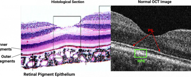
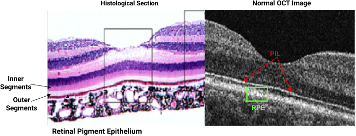
References: 1. Photoreceptor Layer in Central Retinal Vein Occlusion. The Journal of Retinal and Vitreous Diseases; 2008:28(10): 1502-1508 2. Sherman J., Yannuzzi LA., et al., Photoreceptor Integrity Line as Revealed by Spectral Domain OCT. New York 2009; available at lulu.com 3. Vedantham V., Agrawal D. Premacular Hemorrhage Associated with Arteriovenous Communications of the Retina

AV Malformation (AVM) – Page 22 of 222021-11-30T22:25:14+00:00

AV Malformation (AVM) – Page 20 of 22

Jerome Sherman, OD, FAAO Jerome Sherman, O.D., is perhaps optometry's most prolific writer, publishing over 650 clinical articles, research manuscripts, book chapters and two CDs. He is senior author of three books that were published in 2007, and has delivered over three thousand lectures both nationally and internationally. He has served as a contributing

AV Malformation (AVM) – Page 20 of 222021-11-30T22:24:52+00:00

AV Malformation (AVM) – Page 18 of 22

Comments and Conclusions Con’t Arteriovenous communications of the retina can be classified into three groups4: - Group 1-The retina shows interposition of an abnormal capillary plexus between the major vessels, mostly without visual symptoms. - Group 2-The retina reveals direct arteriovenous communications without interposition of capillaries. Patients have few visual symptoms, In addition, associated cerebral

AV Malformation (AVM) – Page 18 of 222021-11-30T22:24:38+00:00

AV Malformation (AVM) – Page 17 of 22

Comments and Conclusions Arteriovenous malformations result from persistence of the embryonic vascular pattern that exists before the arteries, capillaries and veins differentiate during the second month of gestation.4 It is a rare, unilateral nonhereditary disorder with variable visual impairment which depends on the severity of the anomaly. 4 A complete literature search of retinal arteriovenous

AV Malformation (AVM) – Page 17 of 222021-11-30T22:24:32+00:00

AV Malformation (AVM) – Page 16 of 22

Ocular complication of AV Malformation  Although usually non-progressive, retinal arteriovenous anomalies can be associated with the following complications:3 Intraretinal macular hemorrhage Intraretinal hemorrhages outside the macula Neovascular glaucoma Vitreous hemorrhage Central and branch retinal vein occlusion Retinal exudation

AV Malformation (AVM) – Page 16 of 222021-11-30T22:24:24+00:00

AV Malformation (AVM) – Page 15 of 22

Histological Section as Compared to the OCT Image Although the photoreceptor integrity line, or the PIL (defined as the junction between the inner and outer segments) is barely visible in most histological sections, it is highly prominent in normal SD OCTs. The PIL, as shown above should be continuous throughout the entire scan in

AV Malformation (AVM) – Page 15 of 222021-11-30T22:24:17+00:00

AV Malformation (AVM) – Page 14 of 22

The four consecutive OCT scans above reveal the presence of a normal and intact PIL under the foveal pit OS and as well as under the entire macula. The images obtained correlate with BCVA acuity of 20/15. Although both eyes have an epiretinal membrane in the macula, the ERM in the OD is slightly larger

AV Malformation (AVM) – Page 14 of 222021-11-30T22:24:10+00:00

AV Malformation (AVM) – Page 13 of 22

Above are four consecutive horizontal sections through the macula OD. The PIL is near normal and relatively intact superior to the fovea and is absent under the foveal pit. By looking above the letters to be read on the Snellen Chart, the patient is placing the letters to be read on retinal points superior to

AV Malformation (AVM) – Page 13 of 222021-11-30T22:24:03+00:00