

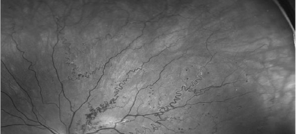
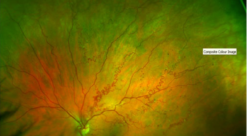
AV Malformation (AVM) – Page 12 of 22
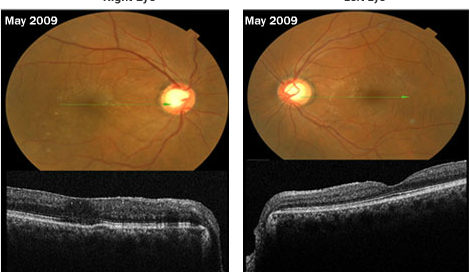
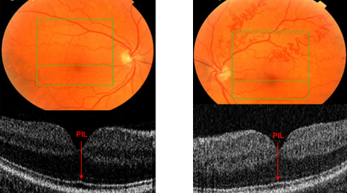
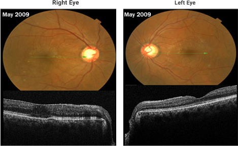
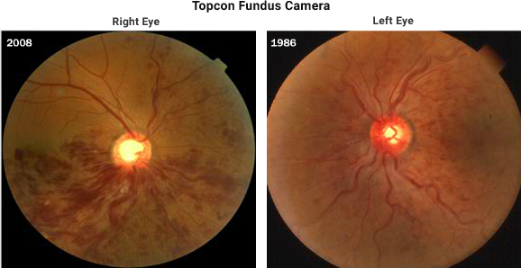
Follow-up of the vein occlusion patient from previous page: The BCVA was 20/30 OD and 20/15 OS. Ophthalmoscopy revealed large cups due to glaucoma OU and an epiretinal membrane 0D>0S. The vein occlusions OD appear to be resolving quite well but the patient is still symptomatic in the right eye. He complains about persistent