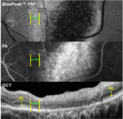
Heidelberg Engineering SPECTRALIS® BluePeak™ FAF, FA, and OCT Images OD
Although fluorescein anglography appears normal In the area depicted, the OCT reveals an apparent collapse of the outer retinal layers and PIL. In the same corresponding zone in FAF, an area of subtle hyperautofluorescence is revealed.


