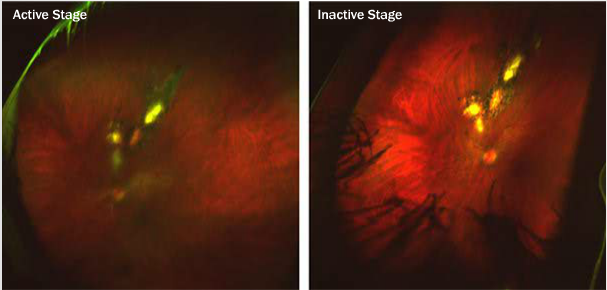
Case 12: Multiple Contiguous Lesions in Retinochoroidal Inflammation Optomap® Color Fundus Images of Left Eye

Case 12: A 35-year-old Hispanic female presented with complaints of increased light sensitivity in her left eye with a history of reoccurring retinal inflammation in the same eye. BCVA measured 20/20 OU. The left eye demonstrated a very hazy view along with four lesions superior to the disc. The three lesions most superior are pigmented but the inferior lesion closest to the disc is not pigmented and appears to be somewhat elevated with ill-defined borders in the left image. The left image was taken before treatment (active stage) and the right image was obtained 2 months after treatment (inactive stage).


