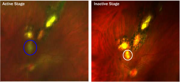
Case 12: Optomap® Fundus Image Comparison of Left Eye

The three pigmented lesions are quite typical of old, inactive retinochoroiditis scars due to toxoplasmosis. In contrast, the lesion closest to the disc with hazy borders without pigment in the left image (see blue circle) is typical of an active toxoplasmosis lesion. Reactivations of toxoplasmosis are most often near or contiguous with old lesions, such as in this case. The patient underwent treatment and within about two months another optomap® image (right image) documented improvement. Note that the lesion closest to the disc in the right image (see white circle) is much better defined than previously. This lesion will pigment within 1 to 2 years. Active toxoplasmosis should be treated if the disc and or macula are threatened. In this case, treatment was indicated because of the proximity of the lesion to the disc.


