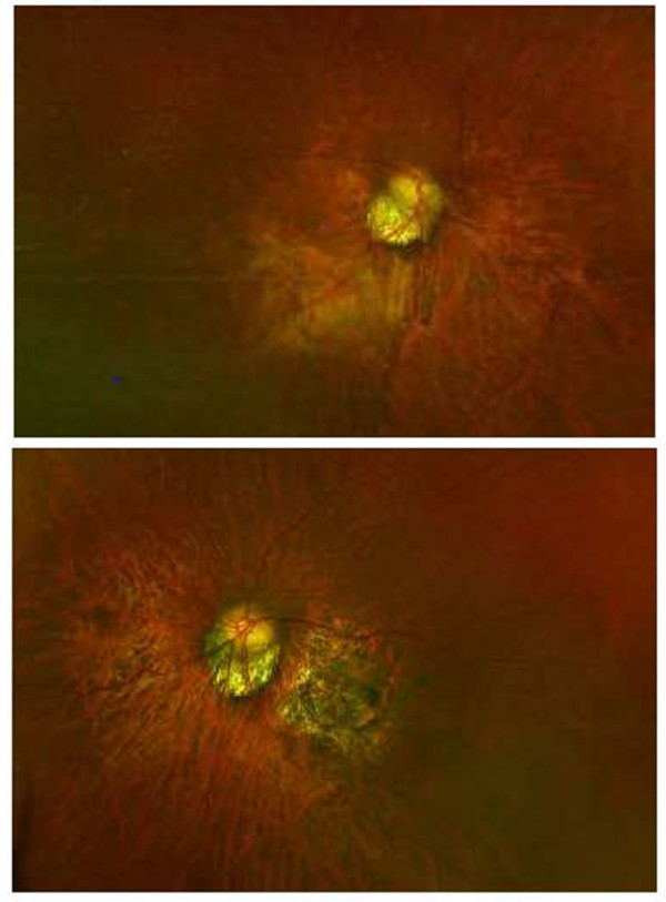
Optomap color fundus photographs

Note the tilted optic nerve heads with posterior staphylomas. Estimated C/D 0.75. Tilting of the optic nerve and posterior staphylomas commonly seen in highly myopic patients make the assessment more challenging and in some cases impossible. The chorioretinal macular scarring in the left eye fundus photograph (bottom) also complicates the diagnosis and management in this patient.


