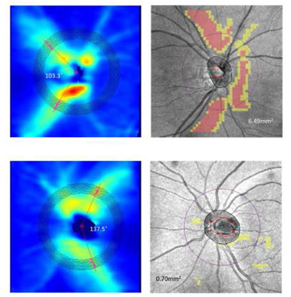
Example of Temporalization of RNFL in Highly Myopic Patients


OCT RNFL heat map and statistical analysis of an eye with spherical error of -6.75 D (top) and -2.00 D (middle). There is a decreased angle between the RNFL bundles showing greater temporalization of RNFL bundles in the more myopic eye.7 The bottom image shows the TSNIT curve on the OCT showing a robust RNFL but shift temporally of the RNFL bundles.
Myopia also causes structural changes within the retina. Due to tilting and increases in axial length, RNFL bundles are shifted temporally. From the work of Don Hood, the superior-temporal and inferior-temporal portions of the optic nerve are the most vulnerable for damage to the optic nerve (superior and inferior vulnerability zones).8 Although these myopic changes are not pathologic, an OCT scan when compared to the normative database, may be flagged as being abnormally thin. Clinicians should look at the TSNIT curve on the OCT and determine if the RNFL is robust but shifted temporally which is commonly seen in highly myopic patients.


