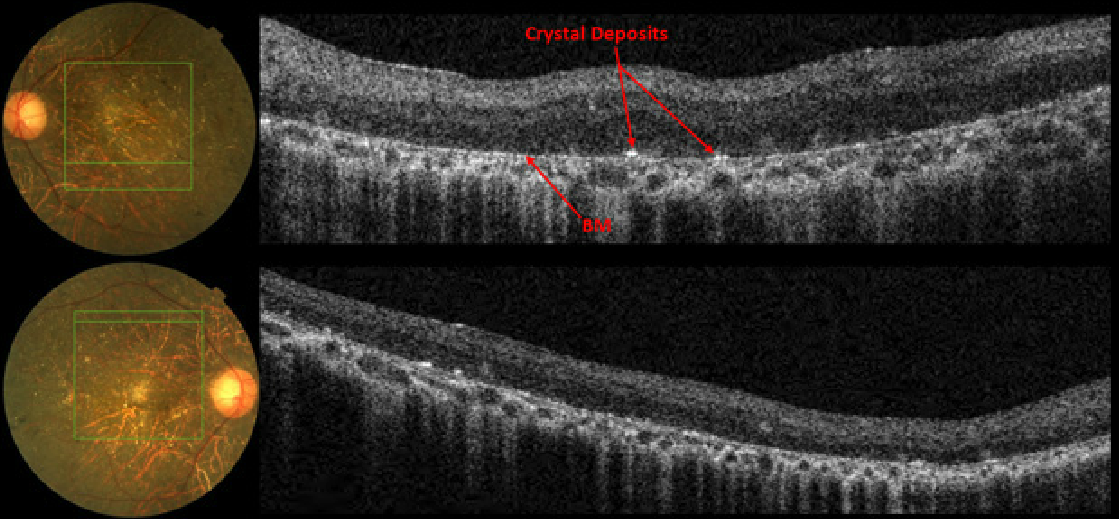
Case 14: Topcon 3D OCT Scan Images

SD-OCT revealed an atrophic retina with no PIL and extremely thin RPE. The yellow crystals appeared as hyperreflective dots on the anterior surface of Bruch’s membrane and more subtle deposits were scattered throughout the other retinal layers. The patient was diagnosed with Bietti’s Crystaline Dystrophy, an autosomal recessive retinal dystrophy, which is characterized by abnormally high levels of triglycerides and cholesterol storage in the body. The yellowish crystals are actually cholesterol/complex lipid inclusions that can be present throughout the body including the retina, cornea, conjunctiva, and choroid. For the complete case report see Retina Revealed: Case #15 – Bietti with Outer Retinal Tubulations


