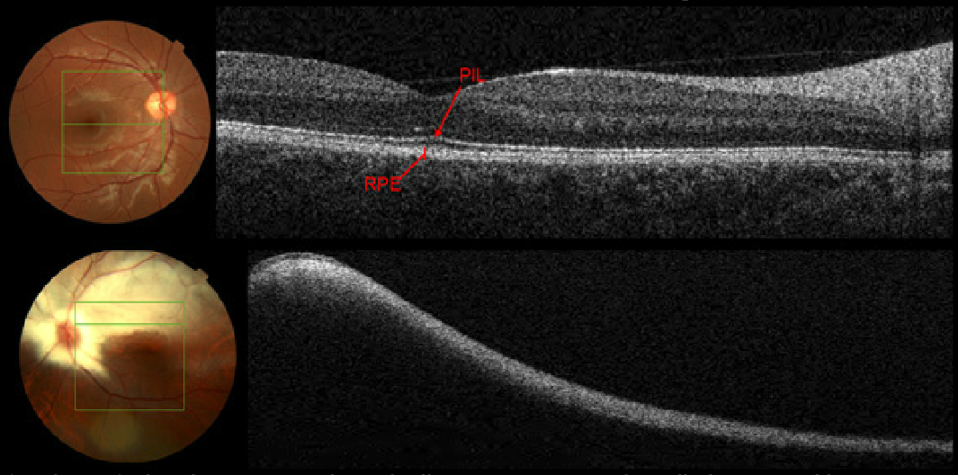
Case 15: Topcon 3D OCT Scan Images

In the right eye (top image), the photoreceptor integrity line appears normal predicting normal best corrected visual acuity. The lower horizontal OCT section above the macula in the left eye reveals a hyperreflective RNFL which is so thick that the underlying structures are “shadowed” or “masked” by the medullated fibers. Myelination should stop at lamina cribrosa (dense aggregation of astrocytes acts as a barrier). Medullated retinal nerve fibers result from the anomalous retinal location of oligodendrocyte-like glial cells as a result of either abnormal migration into the retina prior to the development of the barrier function of lamina cribrosa or abnormal dislocation of the glial cells as a result of a temporary loss of the barrier function of lamina cribrosa.


