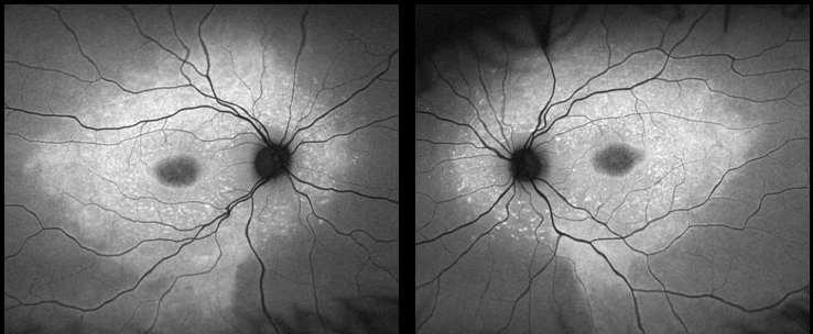
Case 1. (8 yo son) : Optomap® FAF Image OD and OS

Fundus auto-fluorescent (FAF or AF) photos revealed a central hypo-AF oval in each eye. Encircling this was a large hyper-AF zone that extended beyond the arcades. Even brighter hyper-AF spots or flecks were also observed, most marked around the disc and macula of each eye. See Case #RR42 and #RR45 to learn more about AF.


