Case 2 : Topcon 3D OCT Color Fundus Image OU

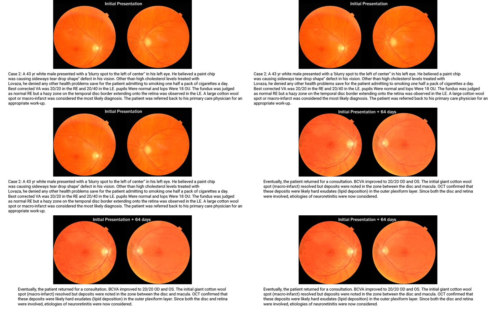
Case 2: A 43 yr white male presented with a “blurry spot to the left of center” in his left eye. He believed a paint chip was causing the “sideways tear drop shape” defect in his vision. Other than high cholesterol levels treated with Lovaza, he denied any other health problems save for the patient admitting to smoking one half a pack of cigarettes a day. Best corrected VA was 20/20 in the RE and 20/40 in the LE. Pupils were normal and IOPs were 18 OU. The fundus was judged as normal RE but a hazy zone on the temporal disc border extending onto the retina was observed in the LE. A large cotton wool spot or macro-infarct was considered the most likely diagnosis. The patient was referred back to his primary care physician for an appropriate work-up.
Eventually, the patient returned for a consultation. BCVA improved to 20/20 OD and OS. The initial giant cotton wool spot (macro-infarct) resolved but deposits were noted in the zone between the disc and macula. OCT confirmed that these deposits were likely hard exudates (lipid deposition) in the outer plexiform layer. Since both the disc and retina were involved, etiologies of neuroretinitis were now considered.


