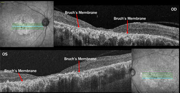
Case 9: Stargardt Disease Cirrus™ HD-OCT Images OU

The blue horizontal section through the fovea in both images reveals the extremely thin macula. Bruch’s membrane is “unmasked” and is well visualized in grey scale images because of the extreme attenuation of the RPE.
For a complete case report on this patient please see: Retina Revealed
Case #14 – Stargardt Plus Glaucoma


