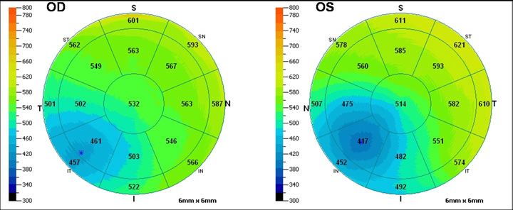
Case 5: iVue Cornea Pachymetry Report OU

In summary: Corneal assessment was also obtained for completion. Corneal pachymetry showed a symmetrical reduction in corneal thickness in both eyes. This is likely due to pellucid marginal degeneration (PMD). A full evaluation of the retina confirmed a retinal degeneration, most likely RP. In advanced RP, ganglion cells are eventually lost as depicted in this case. In early RP, the RNFL is actually thicker than normal.3,4 A literature search has shown that other conditions reported in patients with PMD include retinitis pigmentosa. retinal lattice degeneration, open-angle glaucoma, and several others. However, none of these conditions showed an obvious pathogenic association with PMD.5


