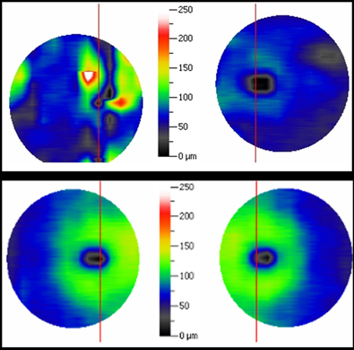
Case 2 (Study Case): iVue GCC Thickness Patient vs. Normal Comparison OU

The patient’s GCC Thickness Map is shown to the left and a normal control is shown below for comparison. The normal control reveals a typical “green donut” in each eye which corresponds to the thickness of GCC. The thin center, which represents the fovea, is normally devoid of ganglion cells. The color scale demonstrates that the GCC is nearly 100 microns in thickness in each eye in this normal patient. In contrast, note that the patient in Case 2 has a GCC which is quite reduced in thickness, more marked in the left eye.


