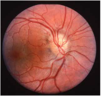
Drusen of the Optic Nerve Head

Drusen bodies of the optic nerve head are often visible ophthalmoscopically and can give the optic nerve head the appearance of a cluster of grapes. Buried drusen, however, are not visible to the eye and are perhaps the most common cause of pseudopapilledema. When drusen are buried, ultrasonography is a useful means by which they may be detected.
Although the nerve head may appear to be elevated on ultrasonography, the key to making the diagnosis is to lower the sensitivity. Since drusen usually contain calcium, they will often continue to reflect sound long after other tissues have become sonolucent at the lower sensitivity settings.
The image to the left is of the optic nerve head of a 12-year old boy with normal visual acuity who presented with what appeared to be blurred disc borders in the right eye.


