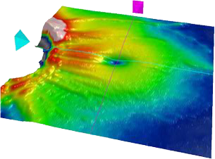
Figure A: ILM-RPE

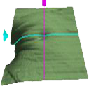
Figure B: Cirrus segmentation of the internal limiting membrane (ILM)

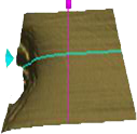
Figure C: Segmentation of the retinal pigment epithelium (RPE)

Horizontal folds are present in the ILM-RPE (Figure A) image and also in the ILM segmentation image (Figure B) but not in the RPE segmentation image (Figure C). Hence, the folds are limited to the ILM.


