

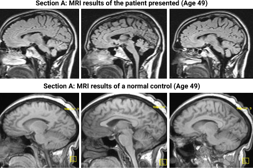
Left column: MRI sagittal 5mm T1 weighted section right cerebellar peduncle level
Middle column: MRI sagittal 5mm T1 weighted midline secction
Right Column: MRI saglttal 5mm T1 weighted left cerebellar peduncle level
The patient scans (A) demonstrate diffuse moderate severity volume loss of the vermis and cerebellar hemispheres as well as of the peduncles and brainstem.