
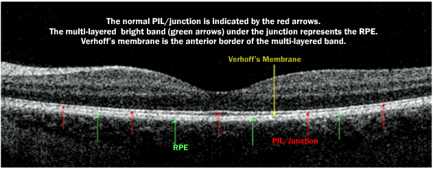
Based upon the integrity of the PIL- also known as the inner/outer (IS/OS) segment junction layer, we can predict patient’s VA. The PIL/junction must be well defined in the fovea in order to achieve good VA. The PIL junction can be used as a “biomarker” for the photoreceptor (both rods and cones) integrity. Verhoff’s membrane is the top of the double or triple layered structure we regard as the RPE. It is not always obvious in imaging, and when visible it appears as the anterior border of the RPE. A disorganized PIL/junction may return to normal under certain conditions.
The PIL as Revealed by SD OCT is available at http://www.lulu.com.
Sponsored by:
