
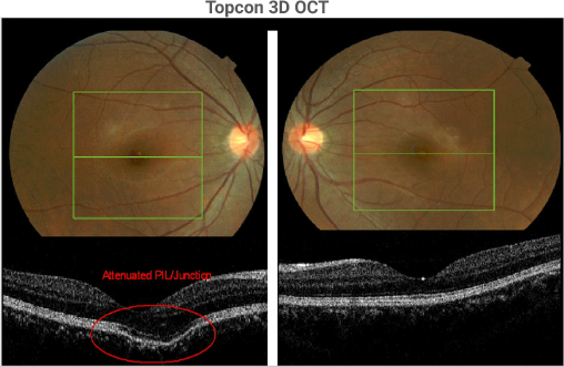
The section through the fovea in the right eye demonstrates the mini-bulge just nasal to the fovea in the image on the left. The PIL/junction (see page 10) is still present but attenuated. The section through the fovea in the left eye is normal. The bulge is likely due to a localized weakness in the sclera. The choroid and the outer retina (including the RPE) appears to dip into the space created by the posterior scleral bulge. The green box is 6 mm x 6 mm and contains 128 horizontal sections.
Sponsored by:
