
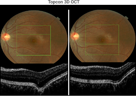
A review of the 128 horizontal sections through the left posterior pole reveals two mini-bulges, one superior to the fovea (above left) and one inferior to the fovea (above right). The most likely interpretation of these two mini-bulges is that they represent very early scleral weakening that could lead to a clinically evident posterior staphloma.
Sponsored by:
