Optos® Fluorescein Angiography

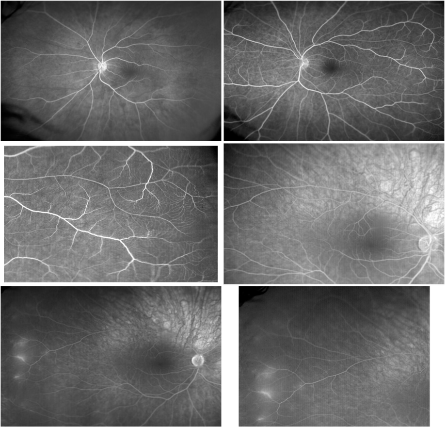
Optomap® fa Dynamic Ultra-widefield Angiography Case 8
It is well recognized that FA reveals select abnormalities that are not visible with ophthalmoscopy or fundus photography. Optos® FA has the advantage of viewing virtually the entire fundus at once and reveals findings typically missed with standard FA.
This is an example of FA in a different patient. Note the peripheral retinal abnormality in the temporal retina OD which represents phlebitis. In this case, the MRI supported the presumed diagnosis of MS. In addition, the FA of the left temporal retina is also not normal. This case as well as Case 4 and 5 will be presented in detail in an upcoming Retina Revealed Issue- stay tuned!


