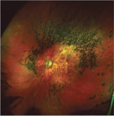
Case 5: Textbook Retinitis Pigmentosa Optomap® Color Fundus Image of Left Eye

Case 5: A 41-year-old female
diagnosed with RP a decade
earlier. BCVA measured 20/25 OIJ.
She complained of her vision
slowing worsening over the last few
years. past medical records
revealed a flat ERG and progressive
field contraction in both eyes.
The optomap® image reveals an
irregular ring of pigment in the mid-
periphery, far denser superiorly
than inferiorly. The retinal vessels
also appeared very attenuated. The
macula also appeared atrophic
with choroidal vasculature and
some sclera showing through the
thin RPE. Note the right eye image
Was virtually identical to the left.


