

Choroidal Detachments
Choroidal detachments present typically as a convex elevation that is attached at the ora serrata with a sonolucent area between the retina-choroid complex and sclera. A choroidal detachment is due to fluid accumulation in the potential space between the choroid and sclera. Unlike a retinal detachment, there is no separation between the retina, RPE and choroid but all three are separated together from the sclera. Choroidal detachments are hence thicker than RDs and appear thicker on B scan. Choroidal detachments are typically in the far peripheral fundus, are convex towards the posterior pole and may be multiple. Usually, there is a recent history of ocular trauma or surgery and low IOPs. Most often, the fluid absorbs without treatment. Unlike RDs which are generally ocular urgencies, choroidal detachments can typically be followed without treatment.
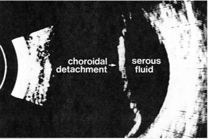
Serous choroidal detachment is depicted in this ultrasonogram. Note the large sonolucent area between the sclera and the retina-choroid complex. Choroid detachments, unlike most retinal detachments, are not attached at the optic nerve head. (Courtesy Biophysic Medical, Inc.)