Case 10: Elevations vs. Excavations

A 49-year-old male with history of a
unilateral congenital optic nerve
abnormality detected five days after
birth and enlarged blind spot in the left
eye. Patient reported that vision in both
eyes is excellent. BCVA measured
20/15+2 OD and 20/15-1 OS.
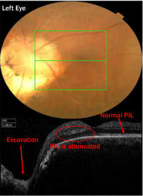
Fundus photo of the anomalous disc
and of the macula, as well as a 6mm x
6mm scan box. The horizontal section
through the macula reveals that the
PIL is normal temporally, becomes
mildly attenuated under the fovea and
eventually disappears towards the
optic nerve head. Note the optic disc
excavation nasal in this scan.
For the full case study go to : Case #8 -“Morning Glory” Disc Coloboma


