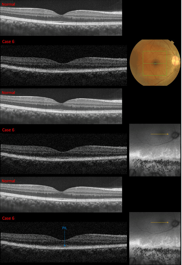
Case 6: Topcon 3D OCT Scan Images OD

A horizontal SD OCT section through the fovea OD reveals a normal PIL under the fovea. However, the PIL appears to thin away from the fovea.

A horizontal SD OCT section through the fovea OD reveals a normal PIL under the fovea. However, the PIL appears to thin away from the fovea.