
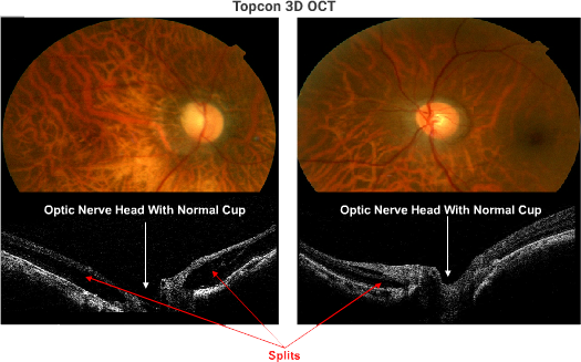
All 128 scans through and around the optic nerve head in the right eye demonstrate a peripapillary retinoschisis (PPRS). Several of these sections demonstrate vitreal-retinal traction which is the probable explanation for the PPRS. A similar RS is present in the left eye which is visualized nasally but not temporally. Similar vitreal-retinal traction is documented in several inferior scans. No optic pits were seen in any of the 128 sections in either eye. Prophylactic vitrectomy is a consideration if central vision is threatened. The dramatic reduction in VA OD could be due to the loss of a PIL in the macula or due to the PPRS or perhaps to a combination of the two abnormalities. Of interest, both are virtually invisible to ophthalmoscopy.
Sponsored by:
