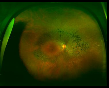
Case 8: Optomap® Color Fundus Image OD

Case 8: A 64 yo HF presented for follow up of possible peri-central RP. Patient reports that she is asymptomatic with no visual complaints at night or day. BCVA was 20/20 OD and OS. Color images revealed extensive peri-central pigment migration greater nasal than temporal in both eyes. Micro perimetry with maia revealed a very minor reduction in sensitivity in the central 10 degrees. Flash ERGs were performed and were in the low normal range under both scoptopic and photopic conditions.


