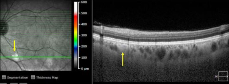
The pathophysiology of these hyper-reflective lesions is not quite understood but there are a few hypotheses:
- Areas of delayed perfusion to the choroid as evidenced with hypofluoresence with indocyanine green.2 Although more recent imaging with optical coherence tomography angiography (OCT-A) reveals these lesions are not associated with flow loss or ischemia in the superficial or deep capillary plexus.3
- Refractile material in the choroid, such as choroidal neurofibromas.4 These choroidal nodules consist of proliferating Schwann cells arranged in concentric rings around an axon, with histological similarities to cutaneous neurofibromata and lisch nodules of the iris.5
When looking specifically through these hyper-reflectant lesions in the choroid with EDI-OCT, they are visualized as areas of choroidal hyper-reflectance with absence of choroidal vasculature (yellow arrows)

These hyper-reflectant choroidal lesions are much more common than you may have realized. In a study by Yasunari et al, these choroidal abnormalities were visible in 100% of patients with NF1, compared to Lisch nodules, only visible in 76% of patients.


