Typical Presentation of CSCR


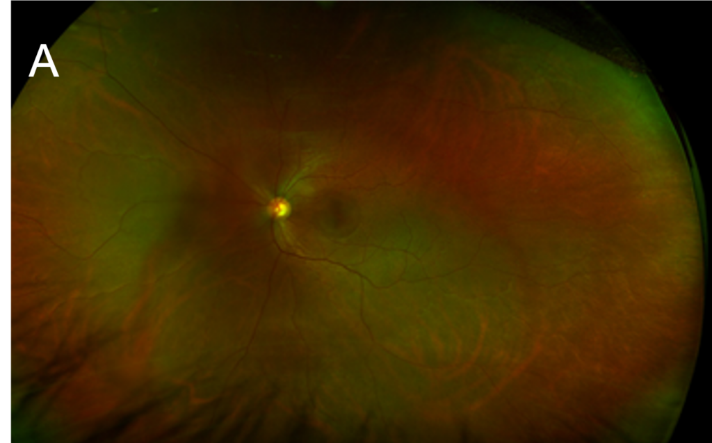
A. Color fundus photograph of left eye without apparent ovoid elevation of retina in the region of CSCR.
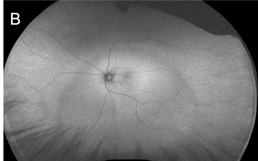
B. Fundus autofluorescence image of left eye with trace hyper autofluorescence demarcation temporal to fovea in the region of CSCR
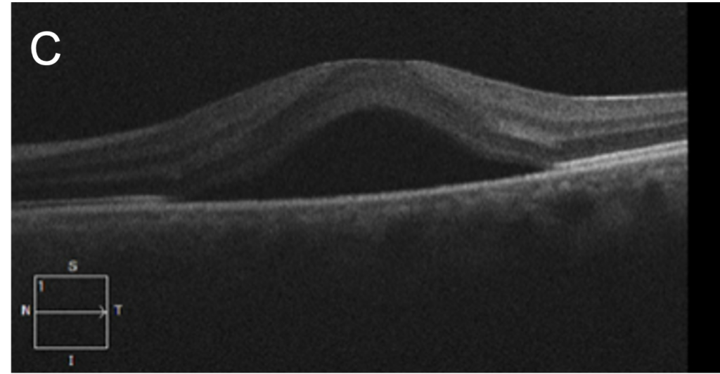
C. OCT scan through left macula showing subretinal fluid and elevation of overlying macula as the typical finding in CSCR.
Note: there is a large circular artifact in pictures A & B that are not CSCR


