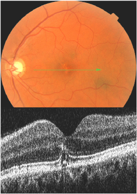
Follow up
- Six months later the patient presented with a one week history of increased distortion on the Amsler grid OS to the left of the central fixation point.
Clinical Findings
- VA OD 20/20 and OS 20/20
- Fundus Exam: Areas of hyperpigmentation and depigmentation in the left macula that appear unchanged from several previous exams.
- Preferential Hyperacuity Perimeter (PHP): Demonstrates normal and unchanged findings.
OCT Image
- In the left eye, several OCT sections through and around the fovea depict several abnormal findings that were not present previously. These include a small zone of hyper-reflectance anterior to the PIL and the penetration of the previous break in the PIL by some tissue that appears to emanate from under the RPE.

See movie presentation on page 1


