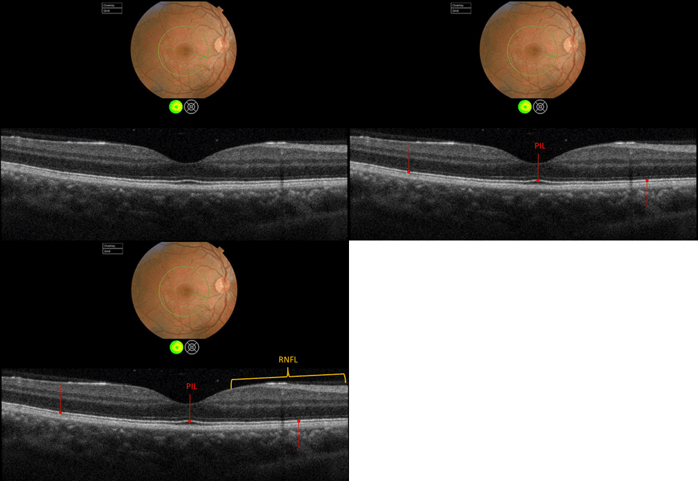
Case 3: Topcon 3D OCT Raster Scan OD

The PIL is intact and normal under the foveal pit and throughout the 6mm scan in this near horizontal section. The RNFL in the papillo-macula bundle also appears intact. Inspection of all the other layers of the retina appear within normal limits as well. Hence, there is no “retinal” explanation for the LP only VA in the right eye.


