Case 10: Suspicious Nevus Topcon 3D OCT Images

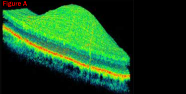
Figure A: The Topcon 3D representation of the lesion’s elevation measuring about 250 microns is depicted on the left. This image is composed of the 128 horizontal slices within the green box.

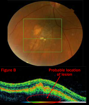
Figure B: One Topcon 3D OCT horizontal slice of the 128 sections contained in the 6×6 mm box is demonstrated to the right. RPE, depicted in red, is elevated due to the underlying drusen and lesion below.
Case 10: A 55-year-old asymptomatic female presented with a lesion in her left eye. BCVA was 20/20 OU. The melanocytic lesion appeared to be a nevus and careful monitoring was recommended. SD OCT is very useful in detecting subtle elevations. Here the elevation was about 1/4 of a mm. For the full case study go to: Retina Revealed Case #7 – Suspicious Nevus


