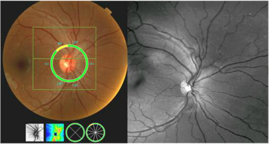
Case 5: Optomap® and RNFL Thickness Analysis OD

Of particular interest, the SD OCT measurement in the zone including the RNFL defect and the area below it are borderline in thickness. This superior temporal zone has an average thickness of 89 µm but the corresponding inferior temporal zone has an average thickness of 147 µm . Is this a normal variant or does it represent early but progressive RNFL loss? Could this be early glaucoma prior to cupping, field loss and pressure elevation? Alternatively, is this indicative of an early non-glaucomatous optic neuropathy? Careful follow-up may shed light on this intriguing case.


