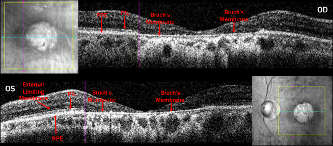
Case 9: Fundus Albi with Cone Dystrophy Cirrus™ HD-OCT Images OU

Case 9: A 64-year-old male who first noticed difficulty with night vision at about age 9 and difficulty reading and seeing faces at age 18 or so. BCVA measured 20/400 OU. OCT sections of the atrophic central lesion reveal a very thin macula and a well visualized Bruch’s membrane. Note the OCT pattern appears somewhat similar to OCT for the Case 5 achromat (see pages 32 and 33).
For a complete case report on this patient please see: Retina Revealed
Case #13 – Fundus Albipunctatus with Cone Dystrophy


