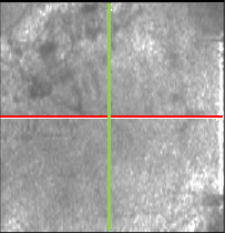
Case 6: Optovue iVue Retinal Cross OCT OD
Retinitis Pigmentosa

Horizontal and vertical iVue sections through the fovea OD of a patient with confirmed retinitis pigmentosa. The PIL is normal in the macula but appears to gradually fade at increased distances from the fovea and appears to “collapse” onto the RPE outside of the macula. This patient has 20/20 VA as predicted by the normal PIL under the fovea. Rods are dramatically reduced as indicated by the absence of the PIL outside of the macula.


