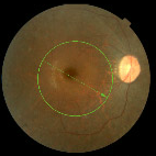
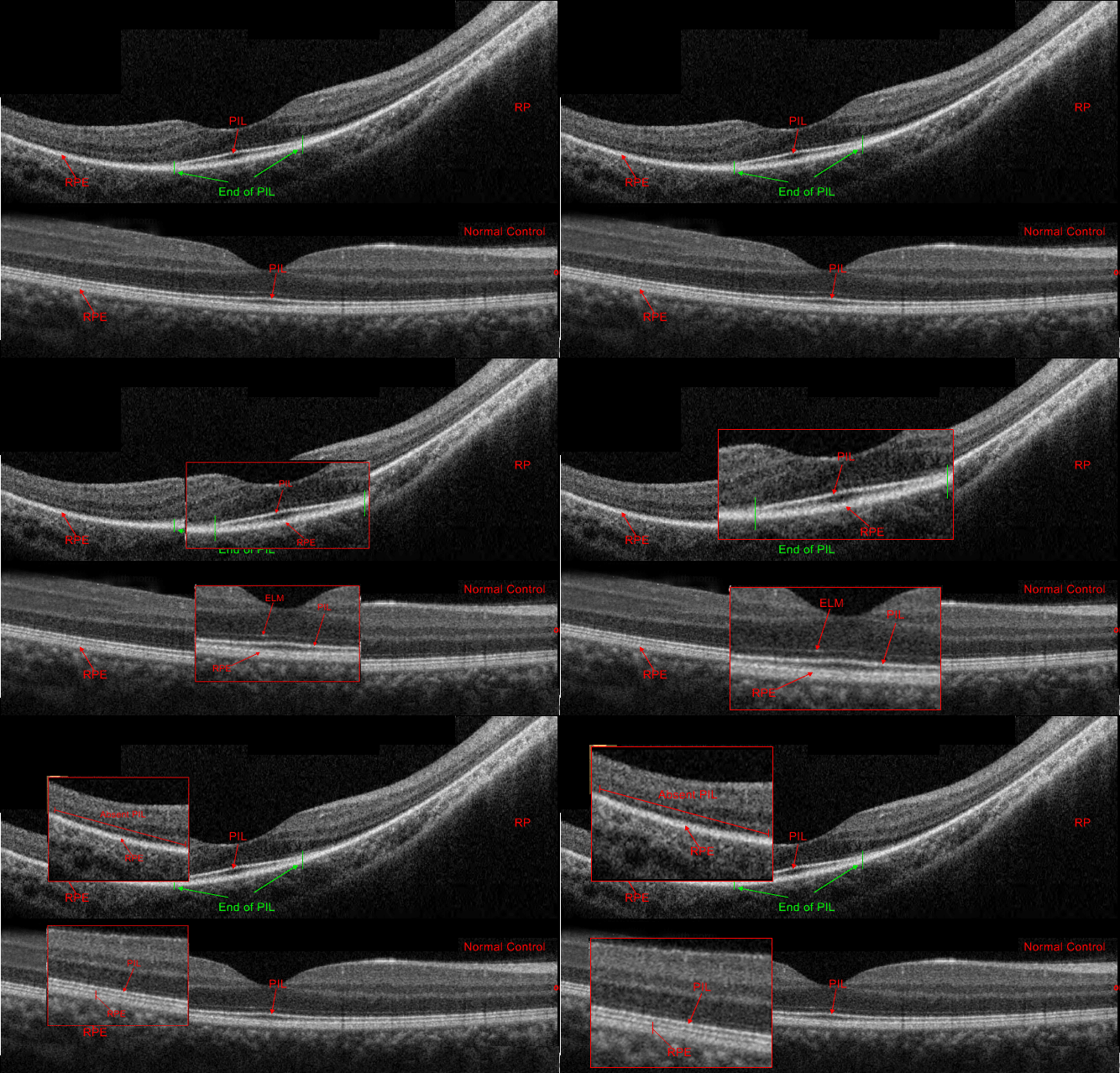
Retinitis Pigmentosa vs. Normal Control Topcon 3D OCT Radial Scan Image of Right Eye


When compared to a normal control, the abnormalities in a RP patient OCT scan are more obvious. Unlike the normal control where the PIL is present throughout, the PIL is absent throughout the scan except a small segment under the fovea in RP patients with normal visual acuity.


