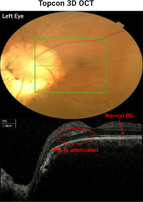
Fundus photo of the anomalous disc and of the macula, as well as a 6mm x 6mm scan box. The horizontal section through the macula reveals that the PIL is normal temporally, becomes mildly attenuated under the fovea and eventually disappears towards the optic nerve head. Note the optic disc excavation nasal in this scan.
Note: In 1970, Kindler first reported an unusual unilateral congenital disc anomaly in 10 patients that resembled the morning glory flower, all with profound unilateral vision reduction.2 Retinoschisis, such as in this case, and retinal detachment have been reported in optic nerve coloboma, MGS and in optic pits, the three conditions that have more recently been grouped into Cavitary Optic Disc Anomalies.1


