

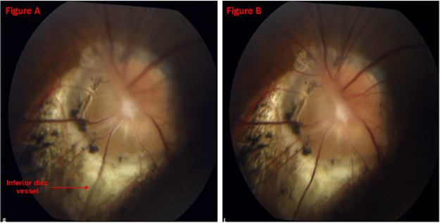
Figure A: The Topcon fundus image is focused on the disc vessels near the cup inferonasally. Note that the large inferior vessel is blurred in the image.
Figure B: In contrast, the fundus image is focused on the large inferior vessel which is somewhat elevated and hence in a different image plane. (Stereopsis may be obtained by converging and fusing the two images)