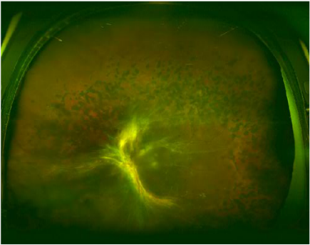
Case 5: Optomap® color fundus image

Case 5: Another patient who has undergone extensive PRP. This patient has traction retinal detachment most obvious inferior to the disc. The fibroglial tissue is tugging on the disc and inferior to the disc in a similar fashion as demonstrated in the SD OCT previously (see pages 4-6).


