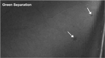
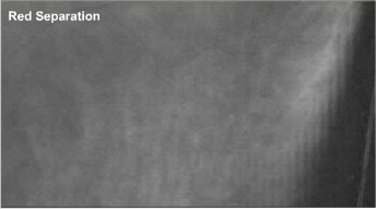
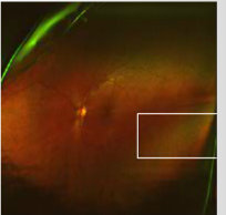
Case 7: Optomap® green and red separation images OS


Separation of the composite color optos® images into red and green channels is helpful in many cases. Subtle findings, such as small retinal hemorrhages at the level of the retina (anterior to the RPE) are most obvious with the green separation and subtle findings at the level of the choroid (posterior to the RPE) are most obvious with the red separation.
Other patients without a reported history of diabetes present with similar small peripheral hemorrhages and when later evaluated, they are diagnosed for the first time with diabetes. Of course, hemorrhages can be due to myriad disorders in addition to diabetes. Peripheral retinal hemorrhages may be the first sign of diabetes and other vascular disorders.


