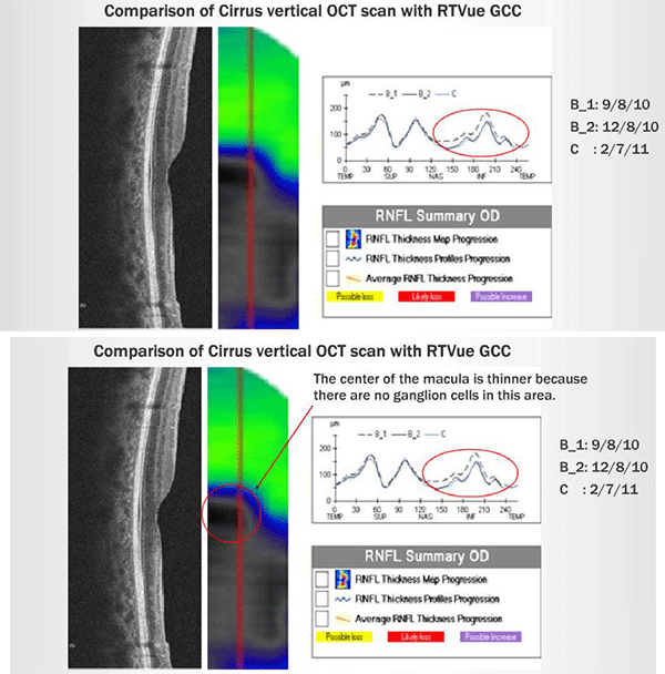
Comparison of Cirrus vertical OCT scan with RTVue GCC

Topcon, Cirrus, and iVue vertical scans through the fovea in the right eye all demonstrate that the inner retina is thinner inferiorly than superiorly as predicted in an occlusion of the inferior branch of the CRA. Shown on the left is the Cirrus vertical section through the fovea and the Optovue GCC. On the right, is the Cirrus Nerve Fiber Layer GPA which does demonstrate some minor progressive loss of the inferior RNFL but this change is not statistically significant (none of the boxes are flagged in yellow, for possible loss, or red, for likely loss).


